Up until now, pregnancy has been a symphony of feelings—flutters, nausea, fatigue, wonder—and a few grainy, early images. But around the halfway mark, something extraordinary happens. You are invited to witness a detailed map of the new life taking shape within you. The anatomy scan at 20 weeks is far more than a routine checkup; it is a pivotal voyage of discovery. This is the moment you move from abstract hope into the realm of intricate, visible reality.
For many, this scan is tinged with a unique duality: breathtaking excitement shadowed by a quiet, vulnerable anxiety known as “scanxiety.” You are about to see your baby in stunning detail, but you are also opening a door to profound knowledge. This guide is your companion for that journey. We will walk through the science and the sentiment of this milestone, demystifying the clinical process while honoring the deep emotional currents that flow through the dimly lit ultrasound room. Consider this your map to the map-making—a way to prepare not just your body, but your heart and mind, for one of pregnancy’s most significant encounters.
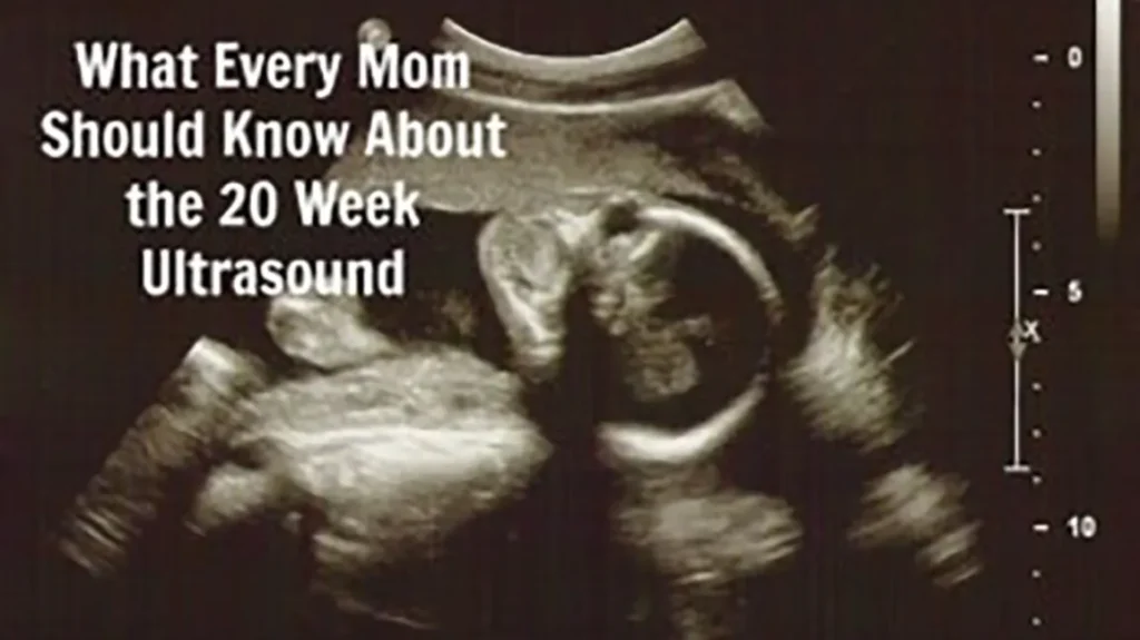
More Than a Peek: The Purpose and Power of the Scan
It’s crucial to reframe this appointment in your mind. This is not a casual “peek at the baby” or merely a gender reveal party (though that can be a joyful part of it). The mid-pregnancy ultrasound is a comprehensive, diagnostic medical examination. Its formal names—the fetal anatomy survey or morphology scan—tell the story: this is a meticulous, system-by-system inventory of your baby’s development.
Think of it as a spacecraft performing a detailed flyby of a new planet, collecting data on every continent, ocean, and atmospheric layer. The sonographer is that skilled navigator, and the transducer is their instrument. The primary goals are threefold:
- To Assess Fetal Anatomy: To confirm that all major organs and body systems are developing typically.
- To Measure Growth: To ensure your baby is growing at an appropriate rate for their gestational age.
- To Evaluate the Environment: To check the placenta, umbilical cord, and amniotic fluid levels—the essential life-support system.
This scan provides a foundational dataset that guides your care for the remainder of the pregnancy. It’s a powerful tool for reassurance and, when necessary, for preparation.
Before You Go: How to Prepare Mind and Body
Preparation is about more than a full bladder. It’s about aligning your practical and emotional selves for the experience.
Practical Logistics
- What to Wear: Opt for comfortable, two-piece clothing (like a shirt and pants or skirt). You’ll need to expose your lower abdomen.
- To Eat or Not to Eat? Yes, eat normally. A active baby is easier to scan. Some clinics even suggest having a small, sugary snack 30 minutes before to encourage movement.
- The Bladder Question: Policies vary. A slightly full bladder can help tilt the uterus for better views early on, but it’s often less critical than at earlier scans. Follow your clinic’s specific instructions.
- Your Partner or Support Person: Their presence is usually encouraged. They are a second set of eyes, a hand to hold, and a crucial part of processing the experience together.
- The Question of “Finding Out”: Decide in advance with your partner if you want to learn the baby’s sex. You can tell the sonographer your preference at the start.
Mental Preparation
- Set an Intention: Beyond “is everything okay?”, what do you need from this? To see a beating heart? To watch your baby move? To simply absorb the reality? A quiet intention can anchor you.
- Acknowledge the Anxiety: “Scanxiety” is real and normal. It’s the fear of the unknown made visual. Allow yourself to feel it, then gently remind yourself that this scan is a tool for knowledge and care, not a test you can fail.
- Discuss the “What Ifs”: Have a gentle conversation with your partner about how you might handle ambiguous news. Hope for the best, but acknowledging the possibility can make you feel more prepared.
The Scan Unfolds: A Moment-by-Moment Journey
The Setting & The Sonographer
You’ll be led into a dimly lit room. The darkness helps the sonographer see the monitor clearly. The centerpiece is the ultrasound machine, with its keyboard, monitors, and the transducer wand. Your guide is the sonographer—a highly trained professional in capturing fetal images. It’s important to know: they are expert imagers, not diagnosticians. They meticulously collect the data; a radiologist or your obstetrician will officially interpret it.
The Process
You’ll lie on the exam table and expose your belly. The sonographer will apply a clear, warm gel (a welcome sensation!) that acts as a conductor for sound waves. Then, the transducer meets your skin.
What to expect during a 20 week ultrasound anatomy scan is a study in focused calm. The sonographer will glide the probe over your belly, pausing, angling, and pressing slightly to get the perfect views. You’ll hear rapid clicks as they freeze images and take measurements with digital calipers. The room may be quiet for long stretches as they concentrate—this is normal and indicates thoroughness, not that something is wrong.
You’ll watch the screen, a window into a hidden world. The images are slices of anatomy, often looking like grainy black-and-white abstractions until the sonographer points out what you’re seeing: “Here’s the profile… there’s the spine… see the four chambers of the heart pumping.”

The Grand Inventory: What the Sonographer is Searching For
This is the heart of the scan—a head-to-toe, inside-out survey. Understanding what does the anatomy scan check for transforms the mysterious clicks and silences into a purposeful narrative.
The Command Center: Brain & Spine
The sonographer will take precise measurements of the fluid-filled spaces (ventricles) in the brain, ensuring they are within normal range. They’ll examine the cerebellum (the part of the brain responsible for coordination) and carefully trace the bony vertebrae of the spine from the neck down to the tailbone, looking for a continuous, intact line to rule out neural tube defects like spina bifida.
The Engine Room: Heart & Circulation
This is a masterpiece of engineering analysis. They will capture the “four-chamber view,” showing the two upper atria and two lower ventricles pumping in rhythm. They’ll then follow the great arteries (the aorta and pulmonary artery) as they exit the heart—the “outflow tracts.” Using color Doppler, they might show you the flow of blood, like a colorful river map, checking for proper direction and velocity.
The Vital Systems: Face, Abdomen & Kidneys
- Face: They will aim for a clear profile view, checking the nasal bone and examining the lips and palate for clefts.
- Abdomen: They’ll locate the stomach bubble (proof the baby is swallowing amniotic fluid), check the diaphragm—the muscle separating chest from abdomen—and examine the liver.
- Kidneys & Bladder: Two kidneys will be visualized, and the presence of a filling and emptying bladder confirms the urinary system is working.
The Infrastructure: Limbs, Bones & Movement
They will measure the femur (thigh bone) and humerus (upper arm bone) to ensure growth is on track. With patience, they’ll try to count fingers and toes. They’ll also observe your baby’s movement—flexing limbs, opening and closing hands, and overall muscle tone. This activity is a vital sign of well-being.
The Living Environment: Placenta, Cord & Fluid
- Placenta: Its location is noted (front/back, top/side). A “low-lying” placenta may need a follow-up scan. Its texture or “grade” is also assessed.
- Umbilical Cord: It should contain three vessels: two arteries and one vein.
- Amniotic Fluid: The sonographer will measure pockets of fluid to ensure there’s not too little (oligohydramnios) or too much (polyhydramnios).
The Aftermath: Understanding Results and Navigating Next Steps
The Immediate Debrief
The sonographer may point out exciting features: “Look, she’s yawning!” or “He’s got your nose!” But they are often limited in what they can interpret. If you ask, “Is everything okay?” they might say, “I’m getting all the images I need,” or “The doctor will review everything.” This is standard protocol, not evasion. Their job is to capture, not diagnose.
The Follow-Up
The images and report are sent to a radiologist or your OB/GYN for official review. How long does the 20 week ultrasound take for results? Often, you’ll hear within a few days, but timelines vary. Your provider will contact you, usually only if there’s a finding to discuss. No news is typically good news.
If a finding requires attention, it often falls into two categories:
- A Clear-Anomaly: This requires a direct conversation with your provider about diagnosis, management, and potential interventions or further testing.
- A “Soft Marker”: This is a subtle finding (like an echogenic intracardiac focus—a bright spot in the heart—or mild pyelectasis—slight kidney dilation) that is often normal but can statistically be associated with a higher chance of a chromosomal condition. It usually warrants a discussion, possibly more genetic counseling or a follow-up scan, but is not a diagnosis in itself.
The Emotional Integration
Leaving the clinic, you may feel elated, drained, or a complex mix. If all looked well, allow yourself a deep sigh of relief—a major milestone is passed. Look at the printed images. Share them. Let the reality sink in. If there are concerns, give yourself grace to feel whatever arises—fear, grief, confusion. Lean on your partner and your provider for clear information and next steps. Knowledge, even hard knowledge, is power.
FAQ: Your Anatomy Scan Anxieties, Addressed
Q: Do I need a full bladder for the 20-week scan?
A: It’s less critical than earlier scans. A partially full bladder can sometimes help initial imaging, but an overly full one can be uncomfortable and actually obstruct views later. Follow your clinic’s specific advice—many now say a comfortable, not full, bladder is fine.
Q: Why won’t the sonographer tell me everything they see?
A: It’s a matter of training and liability. Sonographers are experts in acquiring images, but the final diagnosis and responsibility for interpreting those images lies with the radiologist or your physician. They avoid giving opinions that could be incomplete or cause unnecessary worry.
Q: What’s the difference between 2D, 3D, and 4D ultrasound?
A: 2D is the standard, cross-sectional gray-scale imaging used for all diagnostic measurements. 3D takes multiple 2D slices to create a static, three-dimensional surface image (the baby’s face). 4D is a live 3D video. 3D/4D can be wonderful for bonding but are not typically used for primary diagnosis. Many clinics may offer a quick 3D glimpse at the end of the medical scan if the baby is in a good position.
Q: What are ‘soft markers’ and should I worry about them?
A: A soft marker is an ultrasound finding that is not an abnormality itself but is seen slightly more often in babies with chromosomal conditions. In an otherwise low-risk pregnancy with normal genetic screening, an isolated soft marker is often just a normal variant. Your provider will help you understand the context and if any further action is needed.
Q: Is it normal for the baby to be in a bad position for the scan?
A: Extremely. The baby is an uncooperative, mobile tenant! If key views can’t be obtained, the sonographer may ask you to walk around, drink water, or even return on another day. This is a common, frustrating, but normal part of the process.
Conclusion: The Map is Not the Territory, But It Guides the Way
The anatomy scan at 20 weeks offers an unparalleled glimpse into the hidden architecture of your child. It is a profound intersection of science and soul, where sound waves sketch a portrait of your deepest hopes. Whether you leave floating on a cloud of relief or navigating a new, uncertain path, remember this: the scan does not define your baby. It provides a map—sometimes beautifully clear, sometimes with areas yet to be explored.
This experience, in all its complexity, is a act of witnessing. You have seen the intricate chambers of a beating heart, the graceful curve of a forming spine, the potential of a tiny, waving hand. You have gathered knowledge, and with it, a deeper connection. Carry that connection forward. Let it be the compass for the second half of your journey, reminding you that you are not just carrying a secret, but nurturing a wondrous, detailed, and ever-unfolding life.